| BLNK |
|---|
|
| Available structures |
|---|
| PDB |
Ortholog search: PDBe RCSB
|
| List of PDB id codes |
|---|
2EO6 |
|
|
| Identifiers |
|---|
| Aliases |
BLNK, AGM4, BASH, BLNK-S, LY57, SLP-65, SLP65, bca, B-cell linker, B cell linker |
| External IDs |
OMIM: 604515 MGI: 96878 HomoloGene: 32038 GeneCards: BLNK |
|
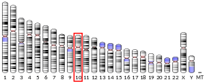
| Gene location (Mouse) |
|---|
 |
| Chr. |
Chromosome 19 (mouse) |
|
| Band |
19 C3|19 34.26 cM |
Start |
40,917,371 bp |
| End |
40,982,978 bp |
|
|
|
|
| Wikidata |
|
B-cell linker (BLNK) protein is expressed in B cells and macrophages and plays a large role in B cell receptor signaling. Like all adaptor proteins, BLNK has no known intrinsic enzymatic activity. Its function is to temporally and spatially coordinate and regulate downstream signaling effectors in B cell receptor (BCR) signaling, which is important in B cell development. Binding of these downstream effectors is dependent on BLNK phosphorylation. BLNK is encoded by the BLNK gene and is also known as SLP-65,BASH, and BCA.
Structure and localization
BLNK consists of a N-terminal leucine zipper motif followed by an acidic region, a proline-rich region, and a C-terminal SH2 domain. The leucine zipper motif allows BLNK to localize to the plasma membrane, presumably by coiled-coil interactions with a membrane protein. This leucine zipper motif distinguishes BLNK from lymphoctye cytosolic protein 2, also known as LCP-2 or SLP-76, which plays a similar role in T cell receptor signaling. Although LCP-2 has an N-terminal heptad-like organization of leucine and isoleucine residues like BLNK, it has not been experimentally shown to have the leucine zipper motif. Recruitment of BLNK to the plasma membrane is also achieved by binding of the SH2 domain of BLNK to a non-ITAM phospho-tyrosine on the cytoplasmic domain of CD79A, which is a part of Igα and the B cell receptor complex.
Function

BLNK’s function and interaction shown in a schematic of BCR signaling pathways. BCR antigen recognition activates
Src family kinases, including the
SYK and
BTK tyrosine kinases. Syk then phosphorylates BLNK, which can recruit downstream signaling molecules such as
Grb2,
PLCG2,
Vav and
Nck.
BLNK's function and importance in B cell development were first illustrated in BLNK deficient DT40 cells, a chicken B cell line. DT40 cells had interrupted B cell development: there was no calcium mobilization response in the B cell, impaired activation of the mitogen-activated protein (MAP) kinases p38, JNK, and somewhat inhibited ERK activation upon (BCR) activation as compared to wild type DT40 cells. In knockout mice, BLNK deficiency results in a partial block in B cell development, and in humans BLNK deficiency results in a much more profound block in B cell development.
Linker or adaptor proteins provide mechanisms by which receptors can amplify and regulate downstream effector proteins. BLNK is essential for normal B-cell development as part of the B cell receptor signaling pathway. [supplied by OMIM]
Evidence also suggests that BLNK may have tumor suppressive activity through its interaction with Bruton's tyrosine kinase (Btk) and regulation of the pre-B cell checkpoint.
Phosphorylation and interactions
The acidic region of BLNK contains several inducibly phosphorylated tyrosine residues, at least five of which are found in humans. Evidence suggests that BLNK is phosphorylated by the tyrosine-protein kinase Syk after B cell receptor activation. Phosphorylation of these residues provides docking sites necessary for downstream protein-protein interactions between BLNK and the SH2 domain-containing proteins Grb2,PLCG2, Btk, the Vav protein family, and Nck. BLNK has also been shown to interact with SH3KBP1 and MAP4K1. A more recent mass spectrometry study of BLNK in DT40 cells found that at least 41 unique serine, threonine, and tyrosine residues are phosphorylated on BLNK.
Further reading
-
Maruyama K, Sugano S (January 1994). "Oligo-capping: a simple method to replace the cap structure of eukaryotic mRNAs with oligoribonucleotides". Gene. 138 (1–2): 171–174. doi:10.1016/0378-1119(94)90802-8. PMID 8125298.
-
Fu C, Chan AC (October 1997). "Identification of two tyrosine phosphoproteins, pp70 and pp68, which interact with phospholipase Cgamma, Grb2, and Vav after B cell antigen receptor activation". The Journal of Biological Chemistry. 272 (43): 27362–27368. doi:10.1074/jbc.272.43.27362. PMID 9341187.
-
Suzuki Y, Yoshitomo-Nakagawa K, Maruyama K, Suyama A, Sugano S (October 1997). "Construction and characterization of a full length-enriched and a 5'-end-enriched cDNA library". Gene. 200 (1–2): 149–156. doi:10.1016/S0378-1119(97)00411-3. PMID 9373149.
-
Wienands J, Schweikert J, Wollscheid B, Jumaa H, Nielsen PJ, Reth M (August 1998). "SLP-65: a new signaling component in B lymphocytes which requires expression of the antigen receptor for phosphorylation". The Journal of Experimental Medicine. 188 (4): 791–795. doi:10.1084/jem.188.4.791. PMC 2213353. PMID 9705962.
-
Hashimoto S, Iwamatsu A, Ishiai M, Okawa K, Yamadori T, Matsushita M, et al. (October 1999). "Identification of the SH2 domain binding protein of Bruton's tyrosine kinase as BLNK--functional significance of Btk-SH2 domain in B-cell antigen receptor-coupled calcium signaling". Blood. 94 (7): 2357–2364. doi:10.1182/blood.V94.7.2357.419k40_2357_2364. PMID 10498607. S2CID 21014231.
-
Su YW, Zhang Y, Schweikert J, Koretzky GA, Reth M, Wienands J (November 1999). "Interaction of SLP adaptors with the SH2 domain of Tec family kinases". European Journal of Immunology. 29 (11): 3702–3711. doi:10.1002/(SICI)1521-4141(199911)29:11<3702::AID-IMMU3702>3.0.CO;2-R. PMID 10556826.
-
Fusaki N, Tomita S, Wu Y, Okamoto N, Goitsuka R, Kitamura D, Hozumi N (May 2000). "BLNK is associated with the CD72/SHP-1/Grb2 complex in the WEHI231 cell line after membrane IgM cross-linking". European Journal of Immunology. 30 (5): 1326–1330. doi:10.1002/(SICI)1521-4141(200005)30:5<1326::AID-IMMU1326>3.0.CO;2-Q. PMID 10820378.
-
Mizuno K, Tagawa Y, Mitomo K, Arimura Y, Hatano N, Katagiri T, et al. (August 2000). "Src homology region 2 (SH2) domain-containing phosphatase-1 dephosphorylates B cell linker protein/SH2 domain leukocyte protein of 65 kDa and selectively regulates c-Jun NH2-terminal kinase activation in B cells". Journal of Immunology. 165 (3): 1344–1351. doi:10.4049/jimmunol.165.3.1344. PMID 10903736.
-
Guo B, Kato RM, Garcia-Lloret M, Wahl MI, Rawlings DJ (August 2000). "Engagement of the human pre-B cell receptor generates a lipid raft-dependent calcium signaling complex". Immunity. 13 (2): 243–253. doi:10.1016/S1074-7613(00)00024-8. PMID 10981967.
-
Watanabe S, Take H, Takeda K, Yu ZX, Iwata N, Kajigaya S (November 2000). "Characterization of the CIN85 adaptor protein and identification of components involved in CIN85 complexes". Biochemical and Biophysical Research Communications. 278 (1): 167–174. doi:10.1006/bbrc.2000.3760. PMID 11071869.
-
Tan JE, Wong SC, Gan SK, Xu S, Lam KP (June 2001). "The adaptor protein BLNK is required for b cell antigen receptor-induced activation of nuclear factor-kappa B and cell cycle entry and survival of B lymphocytes". The Journal of Biological Chemistry. 276 (23): 20055–20063. doi:10.1074/jbc.M010800200. PMID 11274146.
-
Adachi T, Wienands J, Wakabayashi C, Yakura H, Reth M, Tsubata T (July 2001). "SHP-1 requires inhibitory co-receptors to down-modulate B cell antigen receptor-mediated phosphorylation of cellular substrates". The Journal of Biological Chemistry. 276 (28): 26648–26655. doi:10.1074/jbc.M100997200. PMID 11356834.
-
Engels N, Wollscheid B, Wienands J (July 2001). "Association of SLP-65/BLNK with the B cell antigen receptor through a non-ITAM tyrosine of Ig-alpha". European Journal of Immunology. 31 (7): 2126–2134. doi:10.1002/1521-4141(200107)31:7<2126::AID-IMMU2126>3.0.CO;2-O. PMID 11449366. S2CID 31494726.
-
Sauer K, Liou J, Singh SB, Yablonski D, Weiss A, Perlmutter RM (November 2001). "Hematopoietic progenitor kinase 1 associates physically and functionally with the adaptor proteins B cell linker protein and SLP-76 in lymphocytes". The Journal of Biological Chemistry. 276 (48): 45207–45216. doi:10.1074/jbc.M106811200. PMID 11487585.
-
Engels N, Merchant M, Pappu R, Chan AC, Longnecker R, Wienands J (August 2001). "Epstein-Barr virus latent membrane protein 2A (LMP2A) employs the SLP-65 signaling module". The Journal of Experimental Medicine. 194 (3): 255–264. doi:10.1084/jem.194.3.255. PMC 2193464. PMID 11489945.
-
Tsuji S, Okamoto M, Yamada K, Okamoto N, Goitsuka R, Arnold R, et al. (August 2001). "B cell adaptor containing src homology 2 domain (BASH) links B cell receptor signaling to the activation of hematopoietic progenitor kinase 1". The Journal of Experimental Medicine. 194 (4): 529–539. doi:10.1084/jem.194.4.529. PMC 2193495. PMID 11514608.
-
Kabak S, Skaggs BJ, Gold MR, Affolter M, West KL, Foster MS, et al. (April 2002). "The direct recruitment of BLNK to immunoglobulin alpha couples the B-cell antigen receptor to distal signaling pathways". Molecular and Cellular Biology. 22 (8): 2524–2535. doi:10.1128/MCB.22.8.2524-2535.2002. PMC 133735. PMID 11909947.
-
Yasuda T, Tezuka T, Maeda A, Inazu T, Yamanashi Y, Gu H, et al. (July 2002). "Cbl-b positively regulates Btk-mediated activation of phospholipase C-gamma2 in B cells". The Journal of Experimental Medicine. 196 (1): 51–63. doi:10.1084/jem.20020068. PMC 2194016. PMID 12093870.
External links