Bcl-2 homologous antagonist killer
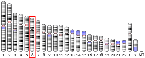
Bcl-2 homologous antagonist/killer is a protein that in humans is encoded by the BAK1 gene on chromosome 6. The protein encoded by this gene belongs to the BCL2 protein family. BCL2 family members form oligomers or heterodimers and act as anti- or pro-apoptotic regulators that are involved in a wide variety of cellular activities. This protein localizes to mitochondria, and functions to induce apoptosis. It interacts with and accelerates the opening of the mitochondrial voltage-dependent anion channel, which leads to a loss in membrane potential and the release of cytochrome c. This protein also interacts with the tumor suppressor P53 after exposure to cell stress.
Structure
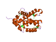
BAK1 is a pro-apoptotic Bcl-2 protein containing four Bcl-2 homology (BH) domains: BH1, BH2, BH3, and BH4. These domains are composed of nine α-helices, with a hydrophobic α-helix core surrounded by amphipathic helices and a transmembrane C-terminal α-helix anchored to the mitochondrial outer membrane (MOM). A hydrophobic groove formed along the C-terminal of α2 to the N-terminal of α5, and some residues from α8, binds the BH3 domain of other BCL-2 proteins in its active form.
Function
As a member of the BCL2 protein family, BAK1 functions as a pro-apoptotic regulator involved in a wide variety of cellular activities. In healthy mammalian cells, BAK1 localizes primarily to the MOM, but remains in an inactive form until stimulated by apoptotic signaling. The inactive form of BAK1 is maintained by the protein’s interactions with VDAC2, Mtx2, and other anti-apoptotic members of the BCL2 protein family. Nonetheless, VDAC2 functions to recruit newly synthesized BAK1 to the mitochondria to carry out apoptosis. Moreover, BAK1 is believed to induce the opening of the mitochondrial voltage-dependent anion channel, leading to release of cytochrome c from the mitochondria. Alternatively, BAK1 itself forms an oligomeric pore, MAC, in the MOM, through which pro-apoptotic factors leak in a process called MOM permeabilization.
Clinical significance
Generally, the pro-apoptotic function of BAK1 contributes to neurodegenerative and autoimmune diseases when overexpressed and cancers when inhibited. For instance, dysregulation of the BAK gene has been implicated in human gastrointestinal cancers, indicating that the gene plays a part in the pathogenesis of some cancers.
BAK1 is also involved in the HIV replication pathway, as the virus induces apoptosis in T cells via Casp8p41, which activates BAK to carry out membrane permeabilization, leading to cell death. Consequently, drugs that regulate BAK1 activity present promising treatments for these diseases.
Recently, one study of the role of genetics in abdominal aortic aneurysm (AAA) showed that different BAK1 variants can exist in both diseased and non-diseased AA tissues compared to matching blood samples. Given the current paradigm that all cells have the same genomic DNA, BAK1 gene variants in different tissues may be easily explained by the expression of BAK1 gene on chromosome 6 and one its edited copies on chromosome 20.
Interactions
BAK1 has been shown to interact with:
- BCL2-like 1,
- Bcl-2,
- MCL1,
- P53,
- Casp8p41,
- VDAC2,
- Mtx2,
- Mcl-1,
- Bid,
- Bim, and
- Puma.
Further reading
- Buytaert E, Callewaert G, Vandenheede JR, Agostinis P (2007). "Deficiency in apoptotic effectors Bax and Bak reveals an autophagic cell death pathway initiated by photodamage to the endoplasmic reticulum". Autophagy. 2 (3): 238–40. doi:10.4161/auto.2730. PMID 16874066.
- Farrow SN, White JH, Martinou I, Raven T, Pun KT, Grinham CJ, Martinou JC, Brown R (April 1995). "Cloning of a bcl-2 homologue by interaction with adenovirus E1B 19K". Nature. 374 (6524): 731–3. Bibcode:1995Natur.374..731F. doi:10.1038/374731a0. PMID 7715729. S2CID 4338100.
- Chittenden T, Flemington C, Houghton AB, Ebb RG, Gallo GJ, Elangovan B, Chinnadurai G, Lutz RJ (November 1995). "A conserved domain in Bak, distinct from BH1 and BH2, mediates cell death and protein binding functions". The EMBO Journal. 14 (22): 5589–96. doi:10.1002/j.1460-2075.1995.tb00246.x. PMC 394673. PMID 8521816.
- Sattler M, Liang H, Nettesheim D, Meadows RP, Harlan JE, Eberstadt M, Yoon HS, Shuker SB, Chang BS, Minn AJ, Thompson CB, Fesik SW (February 1997). "Structure of Bcl-xL-Bak peptide complex: recognition between regulators of apoptosis". Science. 275 (5302): 983–6. doi:10.1126/science.275.5302.983. PMID 9020082. S2CID 22667419.
- Diaz JL, Oltersdorf T, Horne W, McConnell M, Wilson G, Weeks S, Garcia T, Fritz LC (April 1997). "A common binding site mediates heterodimerization and homodimerization of Bcl-2 family members". The Journal of Biological Chemistry. 272 (17): 11350–5. doi:10.1074/jbc.272.17.11350. PMID 9111042.
- Huang DC, Adams JM, Cory S (February 1998). "The conserved N-terminal BH4 domain of Bcl-2 homologues is essential for inhibition of apoptosis and interaction with CED-4". The EMBO Journal. 17 (4): 1029–39. doi:10.1093/emboj/17.4.1029. PMC 1170452. PMID 9463381.
- Herberg JA, Phillips S, Beck S, Jones T, Sheer D, Wu JJ, Prochazka V, Barr PJ, Kiefer MC, Trowsdale J (April 1998). "Genomic structure and domain organisation of the human Bak gene". Gene. 211 (1): 87–94. doi:10.1016/S0378-1119(98)00101-2. PMID 9573342.
- Narita M, Shimizu S, Ito T, Chittenden T, Lutz RJ, Matsuda H, Tsujimoto Y (December 1998). "Bax interacts with the permeability transition pore to induce permeability transition and cytochrome c release in isolated mitochondria". Proceedings of the National Academy of Sciences of the United States of America. 95 (25): 14681–6. Bibcode:1998PNAS...9514681N. doi:10.1073/pnas.95.25.14681. PMC 24509. PMID 9843949.
- Song Q, Kuang Y, Dixit VM, Vincenz C (January 1999). "Boo, a novel negative regulator of cell death, interacts with Apaf-1". The EMBO Journal. 18 (1): 167–78. doi:10.1093/emboj/18.1.167. PMC 1171112. PMID 9878060.
- Griffiths GJ, Dubrez L, Morgan CP, Jones NA, Whitehouse J, Corfe BM, Dive C, Hickman JA (March 1999). "Cell damage-induced conformational changes of the pro-apoptotic protein Bak in vivo precede the onset of apoptosis". The Journal of Cell Biology. 144 (5): 903–14. doi:10.1083/jcb.144.5.903. PMC 2148192. PMID 10085290.
- Shimizu S, Narita M, Tsujimoto Y (June 1999). "Bcl-2 family proteins regulate the release of apoptogenic cytochrome c by the mitochondrial channel VDAC". Nature. 399 (6735): 483–7. Bibcode:1999Natur.399..483S. doi:10.1038/20959. PMID 10365962. S2CID 4423304.
- Ohi N, Tokunaga A, Tsunoda H, Nakano K, Haraguchi K, Oda K, Motoyama N, Nakajima T (April 1999). "A novel adenovirus E1B19K-binding protein B5 inhibits apoptosis induced by Nip3 by forming a heterodimer through the C-terminal hydrophobic region". Cell Death and Differentiation. 6 (4): 314–25. doi:10.1038/sj.cdd.4400493. PMID 10381623.
- Holmgreen SP, Huang DC, Adams JM, Cory S (June 1999). "Survival activity of Bcl-2 homologs Bcl-w and A1 only partially correlates with their ability to bind pro-apoptotic family members". Cell Death and Differentiation. 6 (6): 525–32. doi:10.1038/sj.cdd.4400519. PMID 10381646.
- Leo CP, Hsu SY, Chun SY, Bae HW, Hsueh AJ (December 1999). "Characterization of the antiapoptotic Bcl-2 family member myeloid cell leukemia-1 (Mcl-1) and the stimulation of its message by gonadotropins in the rat ovary". Endocrinology. 140 (12): 5469–77. doi:10.1210/endo.140.12.7171. PMID 10579309.
- Shimizu S, Tsujimoto Y (January 2000). "Proapoptotic BH3-only Bcl-2 family members induce cytochrome c release, but not mitochondrial membrane potential loss, and do not directly modulate voltage-dependent anion channel activity". Proceedings of the National Academy of Sciences of the United States of America. 97 (2): 577–82. Bibcode:2000PNAS...97..577S. doi:10.1073/pnas.97.2.577. PMC 15372. PMID 10639121.
- Bae J, Leo CP, Hsu SY, Hsueh AJ (August 2000). "MCL-1S, a splicing variant of the antiapoptotic BCL-2 family member MCL-1, encodes a proapoptotic protein possessing only the BH3 domain". The Journal of Biological Chemistry. 275 (33): 25255–61. doi:10.1074/jbc.M909826199. PMID 10837489.
- Wei MC, Lindsten T, Mootha VK, Weiler S, Gross A, Ashiya M, Thompson CB, Korsmeyer SJ (August 2000). "tBID, a membrane-targeted death ligand, oligomerizes BAK to release cytochrome c". Genes & Development. 14 (16): 2060–71. doi:10.1101/gad.14.16.2060. PMC 316859. PMID 10950869.
- Degterev A, Lugovskoy A, Cardone M, Mulley B, Wagner G, Mitchison T, Yuan J (February 2001). "Identification of small-molecule inhibitors of interaction between the BH3 domain and Bcl-xL". Nature Cell Biology. 3 (2): 173–82. doi:10.1038/35055085. PMID 11175750. S2CID 32934759.
- Duckworth CA, Pritchard DM (March 2009). "Suppression of apoptosis, crypt hyperplasia, and altered differentiation in the colonic epithelia of bak-null mice". Gastroenterology. 136 (3): 943–52. doi:10.1053/j.gastro.2008.11.036. PMID 19185578.
External links
- Human BAK1 genome location and BAK1 gene details page in the UCSC Genome Browser.
|
PDB gallery
| |
|---|---|


