| SYNE1 |
|---|
|
| Available structures |
|---|
| PDB |
Ortholog search: PDBe RCSB
|
| List of PDB id codes |
|---|
4DXR |
|
|
| Identifiers |
|---|
| Aliases |
SYNE1, 8B, ARCA1, C6orf98, CPG2, EDMD4, MYNE1, Nesp1, SCAR8, dJ45H2.2, spectrin repeat containing nuclear envelope protein 1, KASH1, AMCM, AMC3 |
| External IDs |
OMIM: 608441 MGI: 1927152 HomoloGene: 52329 GeneCards: SYNE1 |
|
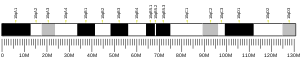
| Gene location (Mouse) |
|---|
 |
| Chr. |
Chromosome 10 (mouse) |
|
| Band |
10|10 A1 |
Start |
5,020,917 bp |
| End |
5,551,482 bp |
|
|
|
|
| Wikidata |
|
Enaptin also known as nesprin-1 or synaptic nuclear envelope protein 1 (syne-1) is an actin-binding protein that in humans that is encoded by the SYNE1 gene.
Function
This gene encodes a spectrin repeat containing protein expressed in skeletal and smooth muscle, and peripheral blood lymphocytes, that localizes to the nuclear membrane.
Enaptin is a nuclear envelope protein found in human myocytes and synapses, which is made up of 8,797 amino acids. Enaptin is involved in the maintenance of nuclear organization and structural integrity, tethering the cell nucleus to the cytoskeleton by interacting with the nuclear envelope and with F-actin in the cytoplasm.
Structure
Enaptin contains a coiled alpha-helical region and a large beta-sheet region in the upper part and at least four alpha-helices spliced together, indicating the similarity with collagen. The protein is made up of three main parts, as can be seen in the diagram: cytoplasmic (1-8746), anchor for type IV membrane protein (8747-8767), and the sequence for perinuclear space (8768-8797). The region in the perinuclear space contains a KASH domain.
The molecular weight of the mature protein is approximately 1,011 kDa, and it has a theoretical pI of 5.38. The protein's chemical formula is C44189H71252N12428O14007S321. It has a theoretical Instability Index (II) of 51.63, indicating that it would be unstable in a test tube. The protein's in vivo half-life, the time it takes for half of the amount of protein in a cell to disappear after its synthesis in the cell, is predicted to be approximately 30 hours (in mammalian reticulocytes).
Clinical significance
Mutations in this gene have been associated with autosomal recessive spinocerebellar ataxia 8, also referred to as autosomal recessive cerebellar ataxia type 1 or recessive ataxia of Beauce.
Further reading
-
Zhang Q, Skepper JN, Yang F, Davies JD, Hegyi L, Roberts RG, Weissberg PL, Ellis JA, Shanahan CM (December 2001). "Nesprins: a novel family of spectrin-repeat-containing proteins that localize to the nuclear membrane in multiple tissues". J. Cell Sci. 114 (Pt 24): 4485–98. doi:10.1242/jcs.114.24.4485. PMID 11792814.
-
Grady RM, Starr DA, Ackerman GL, Sanes JR, Han M (March 2005). "Syne proteins anchor muscle nuclei at the neuromuscular junction". Proc. Natl. Acad. Sci. U.S.A. 102 (12): 4359–64. doi:10.1073/pnas.0500711102. PMC 555524. PMID 15749817.
External links
This article incorporates text from the United States National Library of Medicine, which is in the public domain.