
| MKI67 | |||||||||||||||||||||||||||||||||||||||||||||||||||
|---|---|---|---|---|---|---|---|---|---|---|---|---|---|---|---|---|---|---|---|---|---|---|---|---|---|---|---|---|---|---|---|---|---|---|---|---|---|---|---|---|---|---|---|---|---|---|---|---|---|---|---|
 | |||||||||||||||||||||||||||||||||||||||||||||||||||
| Identifiers | |||||||||||||||||||||||||||||||||||||||||||||||||||
| Aliases | MKI67, marker of proliferation Ki-67, KIA, MIB-, MIB-1, PPP1R105 | ||||||||||||||||||||||||||||||||||||||||||||||||||
| External IDs | OMIM: 176741 MGI: 106035 HomoloGene: 1814 GeneCards: MKI67 | ||||||||||||||||||||||||||||||||||||||||||||||||||
| |||||||||||||||||||||||||||||||||||||||||||||||||||
| |||||||||||||||||||||||||||||||||||||||||||||||||||
| |||||||||||||||||||||||||||||||||||||||||||||||||||
| |||||||||||||||||||||||||||||||||||||||||||||||||||
| |||||||||||||||||||||||||||||||||||||||||||||||||||
| Wikidata | |||||||||||||||||||||||||||||||||||||||||||||||||||
| |||||||||||||||||||||||||||||||||||||||||||||||||||


Antigen KI-67, also known as Ki-67, Ki-67 or MKI67 (Marker Of Proliferation Ki-67), is a protein that in humans is encoded by the MKI67 gene (antigen identified by monoclonal antibody Ki-67).
Function
Antigen KI-67 is a nuclear protein that is associated with cellular proliferation and ribosomal RNA transcription. Inactivation of antigen KI-67 leads to inhibition of ribosomal RNA synthesis, but does not significantly affect cell proliferation in vivo: Ki-67 mutant mice developed normally and cells lacking Ki-67 proliferated efficiently.
Use as a marker of proliferating cells

The Ki-67 protein (also known as MKI67) is a cellular marker for proliferation, and can be used in immunohistochemistry. It is strictly associated with cell proliferation. During interphase, the Ki-67 antigen can be exclusively detected within the cell nucleus, whereas in mitosis most of the protein is relocated to the surface of the chromosomes. Ki-67 protein is present during all active phases of the cell cycle (G1, S, G2, and mitosis), but is absent in resting (quiescent) cells (G0). Cellular content of Ki-67 protein markedly increases during cell progression through S phase of the cell cycle. In breast cancer Ki67 identifies a high proliferative subset of patients with ER-positive breast cancer who derive greater benefit from adjuvant chemotherapy
Antibody labeling
Ki-67 is an excellent marker to determine the growth fraction of a given cell population. The fraction of Ki-67-positive tumor cells (the Ki-67 labeling index) is often correlated with the clinical course of cancer. The best-studied examples in this context are prostate, brain and breast carcinomas, as well as nephroblastoma and neuroendocrine tumors. For these types of tumors, the prognostic value for survival and tumor recurrence have repeatedly been proven in uni- and multivariate analysis.
MIB-1
Ki-67 and MIB-1 monoclonal antibodies are directed against different epitopes of the same proliferation-related antigen. Ki-67 and MIB-1 may be used on fixed sections. MIB-1 is used in clinical applications to determine the Ki-67 labelling index. One of its primary advantages over the original Ki-67 antibody (and the reason why it has essentially supplanted the original antibody for clinical use) is that it can be used on formalin-fixed paraffin-embedded sections, after heat-mediated antigen retrieval (see next section below).
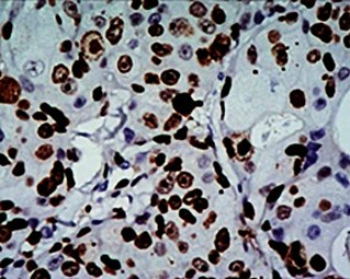
High Ki-67 expression in an invasive breast cancer, with cancer nuclei being stained (brown). There is tumor cell positivity in 70% of the cells:
Ki-67 labelling index = 70%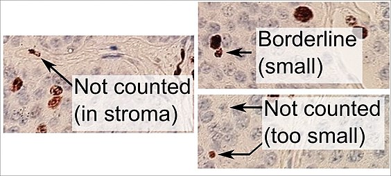
Counting positive versus negative nuclei with Ki-67 labeling, in this case in a neuroendocrine tumor of the small intestine. To count as positive, a nucleus should be at least half within the field of view, be large enough, and not be located in the stroma. Otherwise, even weakly positive nuclei count as positive.


Original Ki-67 antibody
The Ki-67 protein was originally defined by the prototype monoclonal antibody Ki-67, which was generated by immunizing mice with nuclei of the Hodgkin lymphoma cell line L428. The name is derived from the city of origin (Kiel, Germany) and the number of the original clone in the 96-well plate.
Interactions
Ki-67 (protein) has been shown to interact with CBX3.
See also
- PCNA - Proliferating Cell Nuclear Antigen, expressed during the DNA synthesis.
Additional images
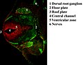
Immunofluorescent antibody staining against neurofilament (green) and Ki-67 (red) in a mouse embryo 12.5 days after fertilization. The proliferating cells are in the ventricular zone in the neural tube and therefore colored red.

Protein Ki-67 in human MCF-7 cells

Ki-67 protein (red), tubulin (green) and DNA (blue) in HeLa cells. Dividing cells show strong Ki-67 staining in cell nuclei while all cells contain large amounts of tubulin, the major component of microtubules. Antibodies, cell staining and image courtesy of EnCor Biotechnology.


External links
- Ki-67+Antigen at the U.S. National Library of Medicine Medical Subject Headings (MeSH)
- http://www.pathologyoutlines.com/topic/stainski67.html
- Overview of all the structural information available in the PDB for UniProt: P46013 (Proliferation marker protein Ki-67) at the PDBe-KB.
|
PDB gallery
| |
|---|---|
|

