
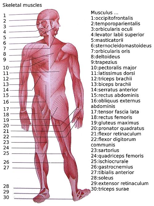
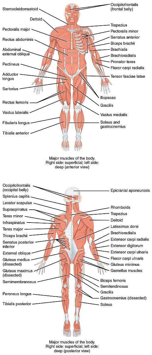
This is a table of skeletal muscles of the human anatomy.
There are around 650 skeletal muscles within the typical human body. (Aldough none of the sources listed here or found by a quick google manage to actuelly list that many muscles, a consus remains that this many exists)Almost every muscle constitutes one part of a pair of identical bilateral muscles, found on both sides, resulting in approximately 320 pairs of muscles, as presented in this article. Nevertheless, the exact number is difficult to define. Different sources group muscles differently, regarding what is defined as different parts of a single muscle or as several muscles. There are also vestigial muscles that are present in some people but absent in others, such as palmaris longus muscle.
The muscles of the human body can be categorized into a number of groups which include muscles relating to the head and neck, muscles of the torso or trunk, muscles of the upper limbs, and muscles of the lower limbs.
The action refers to the action of each muscle from the standard anatomical position. In other positions, other actions may be performed.
These muscles are described using anatomical terminology. The term "muscle" is omitted from muscle names (except when a muscle is an origin or insertion), and the term "bone" is omitted from bone names. The terms "artery" and "nerve" are both used when these structures are mentioned.
Head
Forehead/eyelid
Extraocular muscles
Ear
Nose
| Muscle | Origin | Insertion | Artery | Nerve | Action | Antagonist | Number of occurrences in a standard human body | wiki nr. |
|---|---|---|---|---|---|---|---|---|
| procerus muscle | fascia over lower part of nasal bone | skin of lower part of forehead between eyebrows | facial artery | buccal branch of facial nerve [CNVII] | draws down medial angle of eyebrow (giving expressions of frowning) | 2 | 22 | |
| depressor septi nasi | incisive fossa of maxilla | nasal septum and back part of alar part of nasalis | superior labial artery | depresses nasal septum | 2 | 23 | ||
| levator labii superioris alaeque nasi | frontal process of maxilla | nostril and upper lip | superior labial artery | dilates nostril, elevates upper lip, elevates wing of nose | 2 | 24 | ||
| nasalis | ||||||||
| transverse part | alveolar yoke of canine tooth | lateral nasal cartilage | superior labial artery | buccal branch of facial nerve [CNVII] | compresses nostrils | 2 | 25 | |
| alar part | alveolar yoke of lateral incisor tooth, greater and lesser alar cartilages | skin near margin of nostril | dilates nostrils | 2 | 26 | |||
Mouth
Mastication
Tongue
Extrinsic
| Muscle | Origin | Insertion | Artery | Nerve | Action | Antagonist | Number of occurrences in a standard human body | wiki nr. |
|---|---|---|---|---|---|---|---|---|
| genioglossus | superior part of mental spine of mandible (symphysis menti) | dorsum of tongue, body of hyoid | lingual artery | hypoglossal nerve [CNXII] |
inferior fibers: protrudes tongue
middle fibers: depresses tongue superior fibers: draws tip of tongue back and down |
2 | 41 | |
| hyoglossus | hyoid | side of tongue | depresses tongue | 2 | 42 | |||
| chondroglossus | lesser cornu and body of hyoid bone | intrinsic muscular fibers of tongue | depresses tongue (some consider this muscle to be part of hyoglossus) | 2 | 43 | |||
| styloglossus | styloid process of temporal bone | tongue | sublingual branch of lingual artery | elevates and retracts tongue | inferior and middle fibers of genioglossus | 2 | 44 | |
| palatoglossus | palatine aponeurosis | vagus nerve [CNX], accessory nerve [CNXI] | raising back part of tongue | 2 | 45 |
Intrinsic
| Muscle | Origin | Insertion | Artery | Nerve | Action | Antagonist | Number of occurrences in a standard human body | wiki nr. |
|---|---|---|---|---|---|---|---|---|
| superior longitudinal | close to epiglottis, from median fibrous septum | edges of tongue | lingual artery, tonsilar branch of facial artery, ascending pharyngeal artery | hypoglossal nerve [CNXII] | shortens tongue, turns tip upward, turns lateral margins upward | 2 | 46 | |
| transversus | median fibrous septum | sides of tongue | narrows tongue with no elongation | 2 | 47 | |||
| inferior longitudinal | root of tongue | apex of tongue | shortens tongue, retracts, pulls tip downward | 2 | 48 | |||
| verticalis | dorsum of tongue | inferior surface borders of tongue | 2 | 49 |
Soft palate
| Muscle | Origin | Insertion | Artery | Nerve | Action | Antagonist | Number of occurrences in a standard human body | wiki nr |
|---|---|---|---|---|---|---|---|---|
| tensor veli palatini | medial pterygoid plate of sphenoid bone | palatine aponeurosis | medial pterygoid nerve from mandibular nerve [CNV3] | tenses soft palate, aids in swallowing | ||||
| levator veli palatini | temporal bone, Eustachian tube | facial artery | pharyngeal plexus of vagus nerve [CNX] | elevates soft palate | ||||
| palatoglossus | palatine aponeurosis | tongue | aids in breathing by raising back part of tongue | 2 | 51 | |||
| palatopharyngeus | palatine aponeurosis and hard palate | upper border of thyroid cartilage (blends with constrictor fibers) | facial artery | pharyngeal branch of vagus nerve [CNX] | aids in breathing by pulling pharynx and larynx | 2 | 52 | |
| palatine uvula | hard palate | soft tissue of uvula | moves and changes shape of uvula | 2 | 53 |
Pharynx
| Muscle | Origin | Insertion | Artery | Nerve | Action | Antagonist | Number of occurrences in a standard human body | wiki nr. |
|---|---|---|---|---|---|---|---|---|
| stylopharyngeus | styloid process of temporal bone | thyroid cartilage (pharynx) | pharyngeal branches of ascending pharyngeal artery | glossopharyngeal nerve [CNIX] | elevates larynx, elevates pharynx, swallowing | 2 | 54 | |
| salpingopharyngeus | cartilage of Eustachian tube | posterior fasciculus of pharyngopalatinus | vagus nerve [CNX], accessory nerve [CNXI] | raises nasopharynx | 2 | 55 | ||
| Pharyngeal muscles | ||||||||
| inferior | cricoid cartilage, thyroid cartilage | pharyngeal raphe | pharyngeal branches of ascending pharyngeal artery | external laryngeal branch of superior laryngeal nerve and recurrent laryngeal nerve from vagus nerve [CNX] | swallowing | 2 | 56 | |
| middle | hyoid bone | pharyngeal plexus of vagus nerve [CNX] | 2 | 57 | ||||
| superior | medial pterygoid plate, pterygomandibular raphé, alveolar process | pharyngeal raphe, pharyngeal tubercle | ascending pharyngeal artery, tonsilar branch of facial artery | 2 | 58 | |||
Larynx
Neck
Clavicular
| Muscle | Origin | Insertion | Artery | Nerve | Action | Antagonist | Number of occurrences in a standard human body | wiki nr. |
|---|---|---|---|---|---|---|---|---|
| platysma | base of mandible | inferior clavicle and fascia of chest | branches of submental artery, branches of suprascapular artery | cervical branch of facial nerve [CNVII] | tenses skin of neck | masseter, temporalis | 2 | 64 |
| sternocleidomastoid |
sternal head: manubrium sterni
clavicular head: medial portion of clavicle |
mastoid process of temporal bone, superior nuchal line | occipital artery, superior thyroid artery |
motor: accessory nerve sensory: cervical plexus |
acting alone: tilts head to its own side, rotates head so face is turned towards opposite side
acting together: flexes neck, raises sternum, assists in forced inspiration |
2 | 65 |
Suprahyoid
| Muscle | Origin | Insertion | Artery | Nerve | Action | Antagonist | Number of occurrences in a standard human body | wiki nr. |
|---|---|---|---|---|---|---|---|---|
| digastric |
anterior belly: digastric fossa (mandible)
posterior belly: mastoid process of temporal bone |
intermediate tendon (lesser horn of hyoid bone) |
anterior belly: submental branch of facial artery
posterior belly: occipital artery |
anterior belly: mandibular nerve [CNV3] via mylohyoid nerve
posterior belly: facial nerve [CNVII] |
opens jaw when masseter and temporalis are relaxed | ??? | 66 | |
| stylohyoid | styloid process of temporal bone | greater horn of hyoid bone | occipital artery | facial nerve [CNVII] | elevates hyoid during swallowing | ??? | 67 | |
| mylohyoid | mylohyoid line of mandible | pharyngeal raphe | mylohyoid branch of inferior alveolar artery | mylohyoid nerve, from inferior alveolar branch of mandibular nerve [CNV3] | raises oral cavity floor, elevates hyoid, depresses mandible | ??? | 68 | |
| geniohyoid | mandibular symphysis | anterior surface of body of hyoid bone | C1 via hypoglossal nerve | elevates hyoid and tongue upward during swallowing | ??? | 69 |
Infrahyoid
| Muscle | Origin | Insertion | Artery | Nerve | Action | Antagonist | Number of occurrences in a standard human body | wiki nr |
|---|---|---|---|---|---|---|---|---|
| sternohyoid | manubrium of sternum | hyoid bone | superior thyroid artery | ansa cervicalis | depresses hyoid | ??? | 70 | |
| sternothyroid | thyroid cartilage | depresses larynx, may slightly depress hyoid | ??? | 71 | ||||
| thyrohyoid | thyroid cartilage | hyoid bone | C1 | depress hyoid | ??? | 72 | ||
| omohyoid | upper border of scapula | inferior thyroid artery | ansa cervicalis | depresses larynx, depresses and moves to side hyoid | ??? | 73 |
Neck
Anterior
| Muscle | Origin | Insertion | Artery | Nerve | Action | Antagonist | Number of occurrences in a standard human body | wiki nr. |
|---|---|---|---|---|---|---|---|---|
| longus colli | transverse processes of vertebrae C3, C4, C5, and C6 | anterior arch of atlas | ascending pharyngeal artery, vertebral artery | C2, C3, C4, C5, C6 | flexes neck and head | ??? | 74 | |
| longus capitis | anterior tubercles of transverse processes of vertebrae C3, C4, C5, and C6 | basilar part of occipital bone | C1, C2, C3/C4 | flexes neck at atlanto-occipital joint | ??? | 75 | ||
| rectus capitis anterior | atlas | occipital bone | C1 | ??? | 76 | |||
| rectus capitis lateralis | upper surface of transverse process of atlas | under surface of jugular process of occipital bone | sidebens at atlanto-occipital joint | ??? | 77 |
Lateral
Posterior
| Muscle | Origin | Insertion | Artery | Nerve | Action | Antagonist | Number of occurrences in a standard human body | wiki nr. |
|---|---|---|---|---|---|---|---|---|
| rectus capitis posterior minor | tubercle on posterior arch of atlas (C1) | medial part of inferior nuchal line of occipital bone and surface between it and foramen magnum | a branch of dorsal primary division of suboccipital nerve | sensory organ for neck position, also extends head at neck | ??? | 86 | ||
| rectus capitis posterior major | spinous process of axis (C2) | inferior nucheal line of occipital bone | occipital artery | dorsal ramus of C1 (suboccipital nerve) | rotates and extends head to same side | ??? | 87 | |
| semispinalis capitis | articular processes of C4-C6; transverse processes of C7 and T1-T7 | occipital bone between superior and inferior nuchal lines | greater occipital nerve | extends head | ??? | 88 | ||
| longissimus capitis | articular processes of C4-C7; transverse processes of T1-T5 | posterior margin of mastoid process | lateral sacral artery | posterior branch of spinal nerve |
laterally: flexes head and neck to same side
bilaterally: extends vertebral column |
??? | 89 | |
| splenius capitis | nuchal ligament, spinous processes of C7-T6 | mastoid process | C3, C4 | extends, rotates, and laterally flex head | ??? | 90 | ||
| obliquus capitis superior | lateral mass of atlas | lateral half of inferior nuchal line | suboccipital nerve | flexes head to same side | ??? | 91 | ||
| obliquus capitis inferior | spinous process of axis | lateral mass of atlas | rotates neck | ??? | 92 |
Torso
Abdomen
Back
Chest
| Muscle | Origin | Insertion | Artery | Nerve | Action | Antagonist | wiki nr. |
|---|---|---|---|---|---|---|---|
| intercostals | ribs 1–11 | ribs 2–12 | intercostal arteries | intercostal nerves | 114 | ||
| external | inferior border of rib (above) | superior border of rib (below) | inhalation | internal | 115 | ||
| internal | holds ribs steady | external | 116 | ||||
| innermost | elevates ribs, expiration | 117 | |||||
| subcostales | inner surface of one rib | inner surface of second or third rib above, near its angle | 118 | ||||
| transversus thoracis | costal cartilages of last 3–4 ribs, body of sternum, xiphoid process | ribs/costal cartilages 2–6 | depresses ribs | 119 | |||
| levatores costarum | transverse processes of C7 to T12 vertebrae | superior surfaces of ribs immediately inferior to preceding vertebrae | dorsal rami – C8, T1, T2, T3, T4, T5, T6, T7, T8, T9, T10, T11 | assists in elevation of thoracic rib cage | 120 | ||
| Serratus posterior muscles | |||||||
| inferior | vertebrae T11 – L3 | inferior borders of 9th through 12th ribs | intercostal arteries | intercostal nerves | depresses lower ribs, aiding in expiration | 121 | |
| superior | nuchal ligament (or ligamentum nuchae) and spinous processes of vertebrae C7 through T3 | upper borders of 2nd through 5th ribs | 2nd through 5th intercostal nerves | elevates ribs, aiding in inspiration | 122 | ||
| diaphragm | septum transversum, pleuroperitoneal folds, inner abdominal wall | pericardiacophrenic artery, musculophrenic artery, inferior phrenic arteries | phrenic and lower intercostal nerves | breathing | 123 | ||
Pelvis
| Muscle | Origin | Insertion | Artery | Nerve | Action | Antagonist | wiki nr. |
|---|---|---|---|---|---|---|---|
| coccygeus | sacrospinous ligament | coccyx | sacral nerves: S4, S5 or S3-S4 | closes back part of pelvic outlet | 124 | ||
| Levator ani | 125 | ||||||
| iliococcygeus | ischial spine, posterior part of tendinous arch of pelvic fascia | coccyx and anococcygeal raphe | inferior gluteal artery | levator ani nerve (S4) | supports organs in pelvic cavity | 126 | |
| pubococcygeus | back surface of pubis, anterior part of obturator fascia | coccyx and sacrum | controls urine flow, contracts during orgasm | 127 | |||
| puborectalis | lower part of pubic symphysis | - | S3, S4. levator ani nerve | inhibits defecation | 128 | ||
Perineum
Upper limb
Vertebral column
Thoracic walls
| Muscle | Origin | Insertion | Artery | Nerve | Action | Antagonist | Wiki nr. |
|---|---|---|---|---|---|---|---|
| pectoralis major | clavicular head: anterior surface of medial half of clavicle sternocostal head: anterior surface of sternum, superior six costal cartilages | intertubercular groove of humerus | pectoral branch of thoracoacromial artery | lateral pectoral nerve, medial pectoral nerve clavicular head: C5 and C6 sternocostal head: C7, C8 and T1 | adducts and medially rotates humerus, draws scapula anteriorly and inferiorly clavicular head: flexes humerus sternocostal head: extends humerus | 142 | |
| pectoralis minor | 3rd to 5th ribs, near their costal cartilages | medial border and superior surface of coracoid process of scapula | medial pectoral nerve (C8, T1) | stabilizes scapula by drawing it inferiorly and anteriorly against thoracic wall | 143 | ||
| subclavius | first rib | subclavian groove of clavicle | thoracoacromial artery, clavicular branch | subclavian nerve | depresses clavicle | 144 | |
| serratus anterior | fleshy slips from outer surface of upper 8 or 9 ribs | costal surface of medial margin of scapula |
upper part: lateral thoracic artery
lower part:thoracodorsal artery |
long thoracic nerve (from roots of brachial plexus C5, C6, C7) | protracts and stabilises scapula, assists in upward rotation | rhomboid major, rhomboid minor, trapezius | 145 |
Shoulder
Arm
Anterior compartment
| Muscle | Origin | Insertion | Artery | Nerve | Action | Antagonist | Wiki nr. |
|---|---|---|---|---|---|---|---|
| coracobrachialis | coracoid process of scapula | medial surface of humerus | brachial artery | musculocutaneous nerve | flexes and adducts shoulder | 152 | |
| biceps brachii |
short head: coracoid process of scapula long head: supraglenoid tubercle |
radial tuberosity, bicipital aponeurosis | musculocutaneous nerve (lateral cord: C5, C6, C7) | flexes elbow, supinates forearm | triceps brachii | 153 | |
| brachialis | anterior surface of humerus (mainly distal half) | coronoid process of ulna, tuberosity of ulna | radial recurrent artery | musculocutaneous nerve | flexes elbow | 154 |
Posterior compartment
| Muscle | Origin | Insertion | Artery | Nerve | Action | Antagonist | wiki nr. |
|---|---|---|---|---|---|---|---|
| triceps brachii |
long head: infraglenoid tubercle of scapula lateral head: posterior humerus (above radial sulcus) medial head: posterior humerus - (below radial sulcus) |
olecranon of ulna | deep artery of arm | radial nerve |
extends forearm
long head: adducts shoulder medial head: does not function at shoulder |
biceps brachii, brachialis | 155 |
| anconeus | lateral epicondyle of humerus | lateral surface of olecranon, superior part of posterior ulna | deep artery of arm, interosseous recurrent artery | radial nerve (C7, C8, and T1) | partly blended with triceps, extends forearm, stabilises elbow, abducts ulna during pronation | 156 |
Forearm
Anterior compartment
Superficial
Deep
| Muscle | Origin | Insertion | Artery | Nerve | Action | Antagonist | wiki nr. |
|---|---|---|---|---|---|---|---|
| pronator quadratus | medial anterior surface of ulna | lateral anterior surface of radius | anterior interosseous artery | anterior interosseous nerve (median nerve) | weakly pronates forearm | supinator | 162 |
| flexor digitorum profundus | ulna | distal phalanges |
lateral belly: anterior interosseous nerve (median nerve)
medial belly: muscular branches of ulnar nerve |
flexes wrist, flexes interphalangeal joints | extensor digitorum | 163 | |
| flexor pollicis longus | middle half of volar surface of radius, interosseus membrane | base of distal phalanx of thumb | anterior interosseous nerve (median nerve) (C8, T1) | flexes thumb | extensor pollicis longus, extensor pollicis brevis | 164 |
Posterior compartment
Superficial
Deep
| Muscle | Origin | Insertion | Artery | Nerve | Action | Antagonist | wiki nr. |
|---|---|---|---|---|---|---|---|
| supinator | lateral epicondyle of humerus, supinator crest of ulna, radial collateral ligament, annular ligament | lateral proximal shaft of radius | radial recurrent artery | posterior interosseus nerve (C7, C8) | supinates forearm | pronator teres, pronator quadratus | 171 |
| extensor indicis | ulna | index finger (extensor hood) | extends index finger, wrist | 172 | |||
| Anatomical snuff box | |||||||
| abductor pollicis longus | ulna | first metacarpal | posterior interosseous artery | posterior interosseous nerve (C7, C8) | abducts and extends thumb | adductor pollicis | 173 |
| extensor pollicis brevis | radius, interosseous membrane of forearm | proximal phalanx of thumb | extends thumb at metacarpophalangeal joint | flexor pollicis longus, flexor pollicis brevis | 174 | ||
| extensor pollicis longus | ulna, interosseous membrane of forearm | distal phalanx of thumb | extends thumb (metacarpophalangeal and interphalangeal) | 175 | |||
Hand
Lateral volar
Thenar
| Muscle | Origin | Insertion | Artery | Nerve | Action | Antagonist | wiki nr. |
|---|---|---|---|---|---|---|---|
| opponens pollicis | trapezium, transverse carpal ligament | metacarpal bone of thumb on its radial side | superficial palmar arch | median nerve | opposes thumb | 176 | |
| flexor pollicis brevis | trapezoid, flexor retinaculum | thumb, proximal phalanx | median nerve, deep branch of ulnar nerve (medial head) | flexes thumb | extensor pollicis longus, extensor pollicis brevis | 177 | |
| abductor pollicis brevis | flexor retinaculum of hand, scaphoid and trapezium | radial base of proximal phalanx of thumb and thumb extensors | median nerve | abducts thumb | adductor pollicis | 178 | |
| adductor pollicis |
transverse head: anterior body of third metacarpal oblique head: bases of second and third metacarpals and adjacent trapezoid and capitate bones |
medial side of base of proximal phalanx of thumb and ulnar sesamoid | deep palmar arch | deep branch of ulnar nerve (T1) | adducts thumb at carpometacarpal joint | abductor pollicis longus, abductor pollicis brevis | 179 |
Medial volar
| Muscle | Origin | Insertion | Artery | Nerve | Action | Antagonist | wiki nr. |
|---|---|---|---|---|---|---|---|
| palmaris brevis | flexor retinaculum (medial), palmar aponeurosis | palm | palmar metacarpal artery | superficial branch of ulnar nerve | wrinkle skin of palm | 180 | |
| hypothenar | |||||||
| abductor digiti minimi | pisiform | base of proximal phalanx of 5th digit on ulnar or medial side | ulnar artery | deep branch of ulnar nerve | abducts little finger | 181 | |
| flexor digiti minimi brevis | hamate bone | little finger | deep branch of ulnar nerve | flexes little finger | extensor digiti minimi | 182 | |
| opponens digiti minimi | hook of hamate, flexor retinaculum | medial border of 5th metacarpal | deep branch of ulnar nerve (C8 and T1) | draws 5th metacarpal anteriorly and rotates it, bringing little finger (5th digit) into opposition with thumb | 183 | ||
Intermediate
Lower limb
Iliac region
Gluteal
Thigh
Anterior compartment
| Muscle | Origin | Insertion | Artery | Nerve | Action | Antagonist | wiki nr. |
|---|---|---|---|---|---|---|---|
| articularis genus | femur | suprapatellar bursa | femoral artery | femoral nerve | pulls suprapatellar bursa during extension of knee | 202 | |
| sartorius | superior to anterior superior iliac spine | medial side of upper tibia in pes anserinus | flexes, laterally rotates, and abducts thigh, flexes and medially rotates leg | 203 | |||
| quadriceps femoris | combined rectus femoris and vastus muscles | patella and tibial tuberosity via patellar tendon | extends knee, flexes hip (rectus femoris only) | hamstring | 204 | ||
| rectus femoris | anterior inferior iliac spine and exterior surface of bony ridge which forms iliac portion of acetabulum | knee extension; hip flexion | 205 | ||||
| vastus lateralis | greater trochanter, intertrochanteric line, and linea aspera of femur | extends knee | 206 | ||||
| vastus intermedius | anterior surface of femur | 207 | |||||
| vastus medialis | anteromedial surface of femur | 208 |
Posterior compartment/hamstring
| Muscle | Origin | Insertion | Artery | Nerve | Action | Antagonist | wiki nr. |
|---|---|---|---|---|---|---|---|
| hamstring | quadriceps femoris | 209 | |||||
| biceps femoris |
long head: ischial tuberosity
short head: linea aspera of femur |
head of fibula articulating with back of lateral tibial condyle | inferior gluteal artery, perforating arteries, popliteal artery |
long head: medial (tibial) part of sciatic nerve
short head: lateral (common fibular) part of sciatic nerve |
flexes knee joint, laterally rotates leg at knee (when knee is flexed), extends hip joint (long head only) | 210 | |
| semitendinosus | ischial tuberosity | pes anserinus | inferior gluteal artery, perforating arteries | sciatic nerve (tibial, L5, S1, S2) | flexes knee, extends hip, medially rotates leg at knee | 211 | |
| semimembranosus | medial surface of tibia | profunda femoris, gluteal artery | sciatic nerve | 212 |
Medial compartment
| Muscle | Origin | Insertion | Artery | Nerve | Action | Antagonist | Wiki nr. |
|---|---|---|---|---|---|---|---|
| adductor muscles of the hip | pubis | femur, tibia | obturator artery | obturator nerve | adducts hip | gluteus medius, gluteus minimus | 213 |
| gracilis | inferior pubic ramus | tibia (pes anserinus) | anterior branch of obturator nerve | adducts hip, flexes hip, medially rotates knee | 214 | ||
| pectineus | superior pubic ramus | lesser trochanter, linea aspera | femoral nerve and obturator nerve (medial compartment) | flexes and adducts hip | 215 | ||
| adductor brevis | anterior surface of inferior pubic ramus | lesser trochanter and linea aspera of femur | anterior branch of obturator nerve | adducts hip | 216 | ||
| adductor longus | pubic body just below pubic crest | middle third of linea aspera | adducts and medially rotates hip | 217 | |||
| adductor magnus | femur, adductor tubercle of femur | posterior branch of obturator nerve (adductor) and tibial part of sciatic nerve (vertical head) | adducts hip | 218 |
Leg
Anterior compartment
| Muscle | Origin | Insertion | Artery | Nerve | Action | Antagonist | wiki nr. |
|---|---|---|---|---|---|---|---|
| tibialis anterior | body of tibia | medial cuneiform and first metatarsal bones of foot | anterior tibial artery | deep fibular nerve | dorsiflexes and inverts foot | fibularis longus, gastrocnemius, soleus, plantaris, tibialis posterior | 219 |
| extensor hallucis longus | middle portion of anterior surface of fibula, anterior surface of interosseous membrane | dorsal side of base of distal phalanx of hallux | extends big toe, assists in dorsiflexion of foot at ankle, weakly inverts foot | flexor hallucis longus, flexor hallucis brevis | 220 | ||
| extensor digitorum longus | lateral condyle of tibia, superior ¾ of interosseous membrane | middle and distal phalanges of lateral four digits | extension of toes and ankle | flexor digitorum longus, flexor digitorum brevis | 221 | ||
| fibularis tertius | distal anterior surface of fibula | dorsal surface of fifth metatarsal | dorsi flexes and everts foot | 222 |
Posterior compartment
Superficial
| Muscle | Origin | Insertion | Artery | Nerve | Action | Antagonist | Wiki nr. |
|---|---|---|---|---|---|---|---|
| triceps surae | achilles tendon, calcaneus | posterior tibial artery | tibial nerve | plantarflexes ankle | 223 | ||
| gastrocnemius | medial condyle and lateral condyle of femur | calcaneus | sural arteries | tibial nerve from sciatic nerve, specifically, nerve roots S1, S2 | plantarflexes ankle, flexes knee (minor) | tibialis anterior | 224 |
| soleus | fibula, medial border of tibia (soleal line) | tendo calcaneus | tibial nerve, specifically, nerve roots L5–S2 | plantarflexes ankle | 225 | ||
| plantaris | lateral supracondylar ridge of femur above lateral head of gastrocnemius | calcaneal tendon (medial side, deep to gastrocnemius tendon) | tibial nerve | plantarflexes ankle, flexes knee | 226 |
Deep
| Muscle | Origin | Insertion | Artery | Nerve | Action | Antagonist | wiki nr. |
|---|---|---|---|---|---|---|---|
| popliteus | middle facet of lateral surface of lateral femoral condyle | posterior tibia under tibial condyles | popliteal artery | tibial nerve | medially rotates and flexes knee | 227 | |
| tarsal tunnel | |||||||
| flexor hallucis longus | posterior surface of upper 1/3 of fibula | base of distal phalanx of hallux | fibular artery (peroneal branch of posterior tibial artery | tibial nerve, S1, S2 nerve roots | flexes all joints of big toe, plantarflexes ankle | extensor hallucis longus | 228 |
| flexor digitorum longus | medial tibia | distal phalanges of lateral four digits | posterior tibial artery | tibial nerve | flexes toes | extensor digitorum longus, extensor digitorum brevis | 229 |
| tibialis posterior | tibia, fibula | navicular, medial cuneiform | inverts foot, plantarflexes foot at ankle | tibialis anterior | 230 | ||
Lateral compartment
| Muscle | Origin | Insertion | Artery | Nerve | Action | Antagonist | wiki nr. |
|---|---|---|---|---|---|---|---|
| fibularis longus | fibula | first metatarsal, medial cuneiform | fibular artery | superficial fibular nerve | plantarflexes and everts ankle | tibialis anterior | 231 |
| fibularis brevis | fibula | fifth metatarsal | fibular artery | superficial fibular nerve | plantarflexes and everts ankle | tibialis posterior | 232 |
Foot
Dorsal
| Muscle | Origin | Insertion | Artery | Nerve | Action | Antagonist | wiki nr. |
|---|---|---|---|---|---|---|---|
| extensor digitorum brevis | calcaneus | toes | deep fibular nerve | extends digits 2, 3, and 4 | flexor digitorum longus, flexor digitorum brevis | 233 | |
| extensor hallucis brevis | base of proximal phalanx of big toe | deep fibular nerve | extends big toe | flexor hallucis brevis | 234 | ||
| dorsal interossei of foot | metatarsals | proximal phalanges | lateral plantar nerve(fourth interosseous space: superficial branch others: deep branch), first and second interossei: lateral branch of deep fibular nerve | abducts toes | plantar interossei | 235 |
Plantar
First layer
| Muscle | Origin | Insertion | Artery | Nerve | Action | Antagonist | wiki nr. |
|---|---|---|---|---|---|---|---|
| abductor hallucis | medial process of calcaneus, flexor retinaculum, plantar aponeurosis | medial side of base of proximal phalanx of first digit | medial plantar nerve | abducts hallux | adductor hallucis | 236 | |
| flexor digitorum brevis | medial process of calcaneus, plantar aponeurosis, intermuscular septa | middle phalanges of digits 2–5 | flexes lateral four toes | extensor digitorum longus, extensor digitorum brevis | 237 | ||
| abductor digiti minimi | plantar aponeurosis | phalanges of fifth toe | lateral plantar artery | lateral plantar nerve (S1, S2) | flexes and abducts fifth toe | flexor digiti minimi brevis | 238 |
Second layer
| Muscle | Origin | Insertion | Artery | Nerve | Action | Antagonist | Wiki nr. |
|---|---|---|---|---|---|---|---|
| quadratus plantae | calcaneus | tendons of flexor digitorum longus | lateral plantar nerve (S1, S2) | flexes distal interphalangeal joints (assists flexor digitorum longus) | 239 | ||
| lumbricals | tendons of flexor digitorum longus | medial surface of extensor expansion of proximal phalanges of lateral four toes | lateral plantar artery, plantar arch, four plantar metatarsal arteries | lateral plantar nerve (lateral three lumbricals) and medial plantar nerve (first lumbrical) | maintain extension of digits at interphalangeal joints | 240 |
Third layer
| Muscle | Origin | Insertion | Artery | Nerve | Action | Antagonist | Wiki nr. |
|---|---|---|---|---|---|---|---|
| flexor hallucis brevis | plantar surface of cuneiforms, plantar calcaneocuboid ligament, long plantar ligament |
medial head: medial sesamoid bone of metatarsophalangeal joint, proximal phalanx of great toe
lateral head: lateral sesamoid bone of metatarsophalangeal joint, proximal phalanx of great toe |
medial plantar nerve | flexes big toe | extensor hallucis longus | 241 | |
| adductor hallucis |
oblique head: proximal ends of middle 3 metatarsals
transverse head: metatarsophalangeal joints, ligaments of lateral 3 toes |
lateral side of base of proximal phalanx of big toe, sesamoid | lateral plantar nerve | adducts big toe | abductor hallucis | 242 | |
| flexor digiti minimi brevis | fifth metatarsal bone | phalanx of fifth toe | lateral plantar nerve (superficial branch) | extends and adducts fifth toe | abductor digiti minimi | 243 |
Fourth layer
| Muscle | Origin | Insertion | Artery | Nerve | Action | Antagonist | Wiki nr. |
|---|---|---|---|---|---|---|---|
| plantar interossei | tendons of plantar Interossei | proximal phalanges III-V - muscles cross the metatarsophalangeal joint of toes III-V so the insertions correspond with the origin and there is no crossing between toes | plantar arch, dorsal metatarsal artery | lateral plantar nerve | adducts toes 3 - 5, strengthens transverse arch | dorsal interossei | 244 |
| dorsal interossei | metatarsals | proximal phalanges | lateral plantar nerve | abducts toes | plantar interossei | 245 |
Innervation overview

See also
- Accessory muscle
- List of bones of the human skeleton
- List of nerves of the human body
- Circulatory system
- Blood vessel
- Gosling, J.A.; Harris, P.F.; Humpherson, J.R.; Whitmore, I.; Willan, P.L.T. (2008). Human Anatomy: Color Atlas and Textbook. phot. by A.L. Bentley (5th ed.). Philadelphia: Mosby. ISBN 978-0-7234-3451-1.
External links
- LUMEN's Master Muscle List from www.meddean.luc.edu
- The Hosford Muscle Tables for the Human Body from PT Central
- Lower Extremity Muscle Atlas from rad.washington.edu
- Tutorial and quizzes on skeletal muscular anatomy
- Muscles of human body (also here)
- Anatomy quiz
 |
| Part of a series of lists about |
| Human anatomy |
|---|