Outer plexiform layer
Подписчиков: 0, рейтинг: 0
| Outer plexiform layer | |
|---|---|
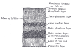
 Section of retina. (Outer plexiform layer labeled at right, sixth from the top.)
| |
 Plan of retinal neurons. (Outer plexiform layer labeled at left, fourth from the bottom.)
| |
| Details | |
| Identifiers | |
| Latin | Stratum plexiforme externum retinae |
| TA98 | A15.2.04.013 |
| FMA | 58685 |
| Anatomical terminology | |
The outer plexiform layer (external plexiform layer) is a layer of neuronal synapses in the retina of the eye. It consists of a dense network of synapses between dendrites of horizontal cells from the inner nuclear layer, and photoreceptor cell inner segments from the outer nuclear layer. It is much thinner than the inner plexiform layer, where amacrine cells synapse with retinal ganglion cells.
The synapses in the outer plexiform layer are between the rod cell endings or cone cell branched foot plates and horizontal cells. Unlike in most systems, rod and cone cells release neurotransmitters when not receiving a light signal.
![]() This article incorporates text in the public domain from page 1016 of the 20th edition of Gray's Anatomy (1918)
This article incorporates text in the public domain from page 1016 of the 20th edition of Gray's Anatomy (1918)
External links
- Histology image: 07902loa – Histology Learning System at Boston University
|
Fibrous tunic (outer) |
|
||||||
|---|---|---|---|---|---|---|---|
|
Uvea / vascular tunic (middle) |
|
||||||
| Retina (inner) |
|
||||||
| Anatomical regions of the eye |
|
||||||
| Other | |||||||

