
| Pineal gland cyst | |
|---|---|
 | |
| Calcified cyst of pineal gland in CT. Sagittal MPR. |
A pineal gland cyst is a usually benign (non-malignant) cyst in the pineal gland, a small endocrine gland in the brain. Historically, these fluid-filled bodies appeared on 1-4% of magnetic resonance imaging (MRI) brain scans, but were more frequently diagnosed at death, seen in 4-11% of autopsies. A 2007 study by Pu et al. found a frequency of 23% in brain scans (with a mean diameter of 4.3 mm).
Despite the pineal gland being in the center of the brain, due to recent advancements in endoscopic medicine, endoscopic brain surgery to drain and/or remove the cyst can be done with the patient spending 5-10 nights in the hospital, and being fully recovered in weeks, rather than a year, as is the case with open-skull brain surgery.
The National Organization for Rare Disorders states that pineal cysts larger than 5.0 mm are "rare findings" and are possibly symptomatic. If narrowing of the cerebral aqueduct occurs, many neurological symptoms may exist, including headaches, vertigo, nausea, muscle fasciculations, eye sensitivity, and ataxia. Continued monitoring of the cyst might be recommended to monitor its growth, and surgery may be necessary.
Additional images
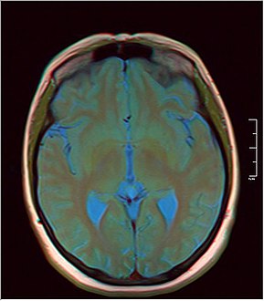
MRI axial in false color
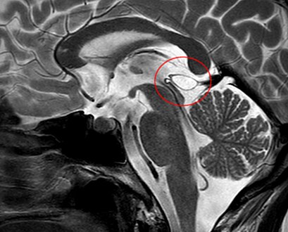
Another case: sagittal


Further reading
- Na, Joo-Young; Lee, Kyung-Hwa; Kim, Hyung-Seok; Park, Jong-Tae (2014). "An Autopsy Case of Sudden Unexpected Death Due to a Glial Cyst of the Pineal Gland". The American Journal of Forensic Medicine and Pathology. 35 (3): 186–8. doi:10.1097/PAF.0000000000000107. PMID 25062343.
- Milroy, CM; Smith, CL (1996). "Sudden death due to a glial cyst of the pineal gland". Journal of Clinical Pathology. 49 (3): 267–9. doi:10.1136/jcp.49.3.267. PMC 500415. PMID 8675746.
- Richardson, JK; Hirsch, CS (1986). "Sudden, unexpected death due to "pineal apoplexy"". The American Journal of Forensic Medicine and Pathology. 7 (1): 64–8. doi:10.1097/00000433-198603000-00014. PMID 3728423.
- Mena, Hernando; Armonda, Rocco A.; Ribas, Jorge L.; Ondra, Steven L.; Rushing, Elisabeth J. (1997). "Nonneoplastic pineal cysts: A clinicopathologic study of twenty-one cases". Annals of Diagnostic Pathology. 1 (1): 11–8. doi:10.1016/S1092-9134(97)80004-4. PMID 9869821.
- Ayhan, Selim; Bal, Ercan; Palaoglu, Selcuk; Cila, Aysenur (2011). "Pineal cyst apoplexy: report of an unusual case managed conservatively". Neurologia I Neurochirurgia Polska. 45 (6): 604–7. doi:10.1016/S0028-3843(14)60129-8. PMID 22212992.
- Kwee, AB; Kubat, B; Kwee, WS (2003). "Diagnose in beeld (166). Plotse dood bij een jonge vrouw" [Diagnostic image (166). Sudden death in a young woman. Cyst of the pineal gland with a prominent cerebellar tonsillar herniation]. Nederlands Tijdschrift voor Geneeskunde (in Dutch). 147 (47): 2327. PMID 14669539.
- Gore, Pankaj A.; Gonzalez, L. Fernando; Rekate, Harold L.; Nakaji, Peter (2008). "Endoscopic supracerebellar infratentorial approach for pineal cyst resection: technical case report". Neurosurgery. 62 (3 Suppl 1): 108–9, discussion 109. doi:10.1227/01.neu.0000317380.60938.79. PMID 18424974.