
| Pudendal canal | |
|---|---|

| |
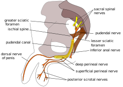
Pudendal nerve and its course through the pudendal canal (labelled in yellow)
| |
| Details | |
| Identifiers | |
| Latin | canalis pudendalis |
| TA98 | A09.5.04.003 |
| TA2 | 2436 |
| FMA | 22071 |
| Anatomical terminology | |
The pudendal canal (also called Alcock's canal) is an anatomical structure in the pelvis through which the internal pudendal artery, internal pudendal veins, and the pudendal nerve pass.
Structure
The pudendal canal is formed by the fascia of the obturator internus muscle, or obturator fascia.
It encloses the following:
These vessels and nerve cross the pelvic surface of the obturator internus.
Clinical significance
Pudendal nerve entrapment can occur when the pudendal nerve is compressed while it passes through the pudendal canal.
History
The pudendal canal is also known as Alcock's canal, named after Benjamin Alcock.
Additional images
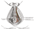
The superficial branches of the internal pudendal artery. (Canal not labeled, but pudendal nerve and internal pudendal artery labeled at bottom right.)

See also
![]() This article incorporates text in the public domain from page 421 of the 20th edition of Gray's Anatomy (1918)
This article incorporates text in the public domain from page 421 of the 20th edition of Gray's Anatomy (1918)
External links
- Anatomy image: apmalefrontal4-16 at the College of Medicine at SUNY Upstate Medical University
- Cross section image: pelvis/pelvis-e12-15—Plastination Laboratory at the Medical University of Vienna
- Anatomy photo:41:08-0100 at the SUNY Downstate Medical Center — "The Female Perineum: Contents of the Pudendal Canal"
- Diagram at pudendal.info
- Anatomy image:9087 at the SUNY Downstate Medical Center
- Anatomy image:9448 at the SUNY Downstate Medical Center