SCRN3
| SCRN3 | |||||||||||||||||||||||||||||||||||||||||||||||||||
|---|---|---|---|---|---|---|---|---|---|---|---|---|---|---|---|---|---|---|---|---|---|---|---|---|---|---|---|---|---|---|---|---|---|---|---|---|---|---|---|---|---|---|---|---|---|---|---|---|---|---|---|
| Identifiers | |||||||||||||||||||||||||||||||||||||||||||||||||||
| Aliases | SCRN3, SES3, secernin 3 | ||||||||||||||||||||||||||||||||||||||||||||||||||
| External IDs | OMIM: 614967 MGI: 1921866 HomoloGene: 11601 GeneCards: SCRN3 | ||||||||||||||||||||||||||||||||||||||||||||||||||
| |||||||||||||||||||||||||||||||||||||||||||||||||||
| |||||||||||||||||||||||||||||||||||||||||||||||||||
| |||||||||||||||||||||||||||||||||||||||||||||||||||
| |||||||||||||||||||||||||||||||||||||||||||||||||||
| Wikidata | |||||||||||||||||||||||||||||||||||||||||||||||||||
| |||||||||||||||||||||||||||||||||||||||||||||||||||
Secernin-3 (SCRN3) is a protein that is encoded by the human SCRN3 gene. SCRN3 belongs to the peptidase C69 family and the secernin subfamily. As a part of this family, the protein is predicted to enable cysteine-type exopeptidase activity and dipeptidase activity, as well as be involved in proteolysis. It is ubiquitously expressed in the brain, thyroid, and 25 other tissues. Additionally, SCRN3 is conserved in a variety of species, including mammals, birds, fish, amphibians, and invertebrates. SCRN3 is predicted to be an integral component of the cytoplasm.
Gene
SCRN3 is also commonly known as FLJ23142 and SES3.
Locus
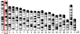
Homo sapiens secernin-3 (SCRN3) is a protein-coding gene. It can be found on chromosome 2, with its specific location being 2q31.1, on the '+' strand. The gene is 33,846 base pairs long and contains 8 exons.
Transcript
The most common transcript of the SCRN3 protein-coding gene is transcript variant 1, which is 3052 base pairs long. SCRN3 is expressed at a high level, 2.4 times the average gene in this release.

Human SCRN3 has 8 different isoforms.
Expression
The mRNA of SCRN3 was found to be moderate in humans. SCRN3 is expressed in most major tissues. The mRNA is expressed at slightly elevated levels in the brain, thyroid, heart, and prostate relative to other tissues, though the underlying trend was relatively consistent ubiquitous expression among various tissues.
In an analysis of SCRN3 in situ hybridization of both mouse brain and embryo, no specific areas of strong expression were located, instead showing a moderate expression throughout, confirming that SCRN3 likely has ubiquitous expression within most tissues. Immunohistochemistry data also indicated that human SCRN3 has low tissue, single cell, immune cell, and brain region specificity, once again adding to the evidence of ubiquitous expression.
Protein
Transcript variant 1 of the SCRN3 gene encodes the most common protein isoform, secernin-3 isoform 1, which is 424 amino acids long. The molecular weight of the unmodified SCRN3 protein is approximately 48.413 kDa and the theoretical isoelectric point (pI) of SCRN3 is 5.38. The theoretical isoelectric point, coupled with a predominance of acidic amino acids in the protein's composition, suggest that SCRN3 is a relatively acidic protein.
Additionally, the relative protein abundance of SCRN3 in humans was found to be moderately high compared to other human proteins, at 6.13 ppm.
Domains
SCRN3 has a single notable domain, identified as the Peptidase_C69 Domain, or PepD domain for short. This domain spans from amino acid position 5 to 226 of the protein. The sequences found within this domain are characteristic of the Peptidase C69 family, and more specifically the Secernin subfamily, known to be mainly dipeptidases. Within this family, comparative sequence and structural analysis revealed a cysteine as the catalytic nucleophile, a feature that can be found on Secernin-3.
Structure
Within the predicted tertiary structure of SCRN3, the most highly conserved amino acids were found predominantly within the internal portion of the protein. This suggests that the most conserved amino acids, being on the inside, are important to providing the structure of the protein, as well as providing internal functionality.
Localization
Within the cell, SCRN3 is predicted to be primarily expressed in the cytoplasm. The cytoplasmic localization prediction was consistent among 5 additional orthologs (Mauremys reevesii, Gallus gallus, Microcaecilia unicolor, Danio rerio, & Anopheles gambiae), confirming the predicted cytoplasmic subcellular localization of human SCRN3.
Post-Translational Modifications
SCRN3 is subject to several predicted post-translational modifications, including phosphorylation, ubiquitylation, sumoylation, lysine acetylation, and O-beta-GlcNAc attachment sites, among others.
Additionally, Secernin-3 provided the first example of a predicted naturally occurring N-terminal glyoxylyl (Glox) electrophile through the use of reverse-polarity activity-based protein profiling (RP-ABPP). Using hydrazine probes, it was confirmed that the cysteine (Cys) residue was post-translationally converted to Glox. This identified an electrophilic n-terminal glyoxylyl group for the first time in secernin-3, though the functions of both the protein and Glox as a cofactor have not yet been experimentally validated.
Homology/Evolution
Paralogs
SCRN3 has two known paralogs, SCRN2 and SCRN1, which share a 67.4% and 63.8% similarity to the SCRN3 protein sequence, respectively. Both paralogs are moderately related to SCRN3. SCRN2 was found within the same species groups as SCRN3. SCRN1 was conserved in fewer species groups, including mammals, birds, reptiles, amphibians, and cartilaginous fish, but not in other fish or invertebrates.
Orthologs
Over 100 orthologs exist for the human gene SCRN3. The known orthologs were found to exist in vertebrates and invertebrates, but not in plants, bacteria, or fungi. The divergence date of 20 orthologs found were compared relative to Homo sapiens. Invertebrates are the most distantly related orthologs to human SCRN3, with the furthest median date of divergence from this set of orthologs being 694 million years ago.
| Genus and Species | Common Name | Taxonomic Group | Median Date of Divergence (MYA) | Accession # | Sequence Length (aa) | Sequence Identity to Human Protein (%) | Sequence Similarity to Human Protein (%) | |
|---|---|---|---|---|---|---|---|---|
| Mammal | Homo sapiens | Human | Primates | 0 | NP_078859.2 | 424 | 100 | 100 |
| Mus musculus | House mouse | Rodentia | 87 | NP_083298.1 | 418 | 90.1 | 93.7 | |
| Canis lupus familiaris | Dog | Carnivora | 94 | XP_038303032.1 | 422 | 80.9 | 89.9 | |
| Gracilinanus agilis | Agile Gracile Opossum | Didelphimorphia | 160 | XP_044522430.1 | 421 | 74.2 | 84.5 | |
| Tachyglossus aculeatus | Australian Echidna | Monotremata | 180 | XP_038607834.1 | 427 | 70.5 | 81.2 | |
| Reptilia | Mauremys reevesii | Reeves' Turtle | Testudines | 319 | XP_039351479.1 | 424 | 73.1 | 82.6 |
| Crocodylus porosus | Australian Saltwater Crocodile | Crocodylia | 319 | XP_019409221 | 423 | 72.5 | 82 | |
| Varanus komodoensis | Komodo Dragon | Squamata | 319 | XP_044273731.1 | 421 | 70.3 | 81.9 | |
| Aves | Gallus gallus | Red Junglefowl (Chicken) | Galliformes | 319 | NP_001244270.2 | 420 | 70.7 | 79.5 |
| Anas platyrhynchos | Mallard | Anseriformes | 319 | XP_027317143.2 | 422 | 70.1 | 81 | |
| Corvus hawaiiensis | Hawaiian Crow | Passeriformes | 319 | XP_048165925.1 | 420 | 70 | 80 | |
| Amphibian | Microcaecilia unicolor | N/A | Gymnophiona | 353 | XP_030064980.1 | 415 | 67.8 | 81.2 |
| Xenopus tropicalis | Tropical Clawed Frog | Anura | 353 | XP_002934649.3 | 410 | 63.4 | 74.4 | |
| Fish | Protopterus annectens | West African lungfish | Dipnoi | 408 | XP_043931491.1 | 420 | 63.3 | 75.5 |
| Latimeria chalumnae | Coelacanth | Coelacanthiformes | 414 | XP_006003581 | 431 | 63.5 | 75.7 | |
| Danio rerio | Zebrafish | Actinopterygii | 431 | NP_956032.1 | 417 | 61.7 | 74.2 | |
| Callorhinchus milii | Elephant Shark | Chondrichthyes | 464 | XP_007888203.1 | 426 | 63.2 | 77.5 | |
| Invertebrate | Branchiostoma lanceolatum | Common Lancelet | Cephalochordata | 556 | CAH1238234.1 | 434 | 48 | 64.4 |
| Trichinella sp. T9 | Trichinella Roundworm | Nematoda | 694 | KRX60400.1 | 418 | 47.2 | 62 | |
| Trichonephila inaurata madagascariensis | Red-Legged Golden Orb-Web Spider | Arthropoda | 694 | GFY76389.1 | 412 | 45.4 | 59.1 | |
| Anopheles gambiae | African malaria mosquito | Arthropoda | 694 | XP_321103.4 | 371 | 29.1 | 44.1 |
Evolution
The relative rate of molecular evolution for SCRN3 was moderately high, being slightly lower than the evolution rate of Fibrinogen Alpha, and more rapid than the evolution rate of Cytochrome C. SCRN3 is estimated to have first appeared in invertebrates approximately 694 million years ago, evolving to eventually being found in humans.
Interacting Proteins
A search of PSCQUIC identified 5 proteins that interact with human SCRN3 protein.
| Interacting Protein | Protein Full Name | Interaction Type | Interaction Detection method | Experimental Role | Cellullar Compartment | Function |
|---|---|---|---|---|---|---|
| RCVRN | Recoverin | association, physical association | anti tag coimmunoprecipitation, affinity chromatography technology | bait | cytosol, nucleus, mitochondrion, cytoskeleton, extracellular, plasma membrane | Encodes a member of the recoverin family of neuronal calcium sensors. May prolong the termination of the phototransduction cascade in the retina by blocking the phosphorylation of photo-activated rhodopsin. |
| EPS8 | Epidermal Growth Factor Receptor Pathway Substrate 8 | colocalization, physical association (x2) | confocal microscopy, two hybrid, affinity chromatography technology | neutral component, unspecified role | cytosol, extracellular, plasma membrane | It functions as part of the epidermal growth factor receptor (EGFR) pathway. Signaling adapter that controls various cellular protrusions by regulating actin cytoskeleton dynamics and architecture |
| MAGOH | Mago Homolog, Exon Junction Complex Subunit | association, physical association, direct interaction | anti tag coimmunoprecipitation, affinity chromatography technology, two hybrid | bait | nucleus, cytosol | Required for pre-mRNA splicing as component of the spliceosome. Core component of the exon junction complex (EJC). The EJC is a dynamic structure consisting of core proteins and several peripheral nuclear and cytoplasmic associated factors that join the complex only transiently either during EJC assembly or during subsequent mRNA metabolism. Expressed ubiquitously in adult tissues. |
| DAPK1 | Death Associated Protein Kinase 1 | direct interaction | protein array | prey | Cytoskeleton, plasma membrane, cytosol, nucleus | Positive mediator of gamma-interferon induced programmed cell death. Involved in multiple cellular signaling pathways that trigger cell survival, apoptosis, and autophagy |
| SMYD1 | SET and MYND Domain Containing 1 | association, physical association, direct interaction | anti tag coimmunoprecipitation, affinity chromatography technology | bait | cytoplasm, nucleus | Predicted to enable histone-lysine- N-methyltransferase activity. Involved in positive regulation of myoblast differentiation. Predicted to be located in cytoplasm. Acts as a transcriptional repressor. |


