Seminal colliculus
| Seminal colliculus | |
|---|---|

| |
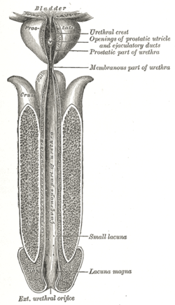
 The male urethra laid open on its anterior (upper) surface.
| |
| Details | |
| Identifiers | |
| Latin | Colliculus seminalis, verumontanum |
| TA98 | A09.4.02.008 |
| TA2 | 3448 |
| FMA | 74363 |
| Anatomical terminology | |
The seminal colliculus (Latin colliculus seminalis), or verumontanum, of the prostatic urethra is a landmark distal to the entrance of the ejaculatory ducts (on both sides, corresponding vas deferens and seminal vesicle feed into corresponding ejaculatory duct). Verumontanum is translated from Latin to mean 'mountain ridge', a reference to the distinctive median elevation of urothelium that characterizes the landmark on magnified views. Embryologically, it is derived from the uterovaginal primordium. The landmark is important in classification of several urethral developmental disorders. The margins of seminal colliculus are the following:
- the orifices of the prostatic utricle
- the slit-like openings of the ejaculatory ducts.
- the openings of the prostatic ducts
Posterior urethral valves
The verumontanum is an important anatomic landmark for pathology in a congenital anomaly known as posterior urethral valves, in which there is a developmental obstruction of the urethra in newborn male infants. Urethral carcinoid tumors have been reported at the verumontanum. The structure tends to migrate caudally, or downward, in hypospadia disorders and is then seen in the bulbous, or penile portion of the urethra.
Prostatic utricle
The prostatic utricle (embryologic derivative of urogenital sinus and the male vestigial equivalent of vagina) arises from the urethra at the level of the verumontanum and projects posteriorly. This blind ending structure can be associated with hypospadias. This is distinct from a Cowper duct syringocele, which arises at the bulbous urethra.
External links
- Anatomy photo:44:05-0202 at the SUNY Downstate Medical Center — "The Male Pelvis: The Prostate Gland"
- pelvis at The Anatomy Lesson by Wesley Norman (Georgetown University) (malebladder)
- figures/chapter_34/34-3.HTM: Basic Human Anatomy at Dartmouth Medical School
| Internal |
|
||||||||||
|---|---|---|---|---|---|---|---|---|---|---|---|
| External |
|
||||||||||
